
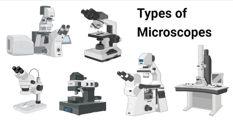
5 Types of Microscopes
There are mainly 5 common types of microscopes as follows:
- Bright-Field or Light Microscope
- Dark Field Microscope
- Phase Contrast Microscope
- Fluorescence Microscope
- Electron Microscope
A good microscope should have three properties:
- Good resolution: Resolution power refers to the ability to produce separate images of closely placed objects so that they can be distinguished as two separate entities. The resolution power of:
- The unaided human eye is about 0.2 mm (200 μm)
- The light microscope is about 0.2 μm
- An electron microscope is about 0.5 nmThe resolution depends on the refractive index. Oil has a higher refractive index than air.
- Good contrast: This can be further improved by staining the specimen.
- Good magnification: This is achieved by the use of concave lenses.
Bright-Field or Light Microscope
The bright-field or light microscope forms a dark image against a brighter background.

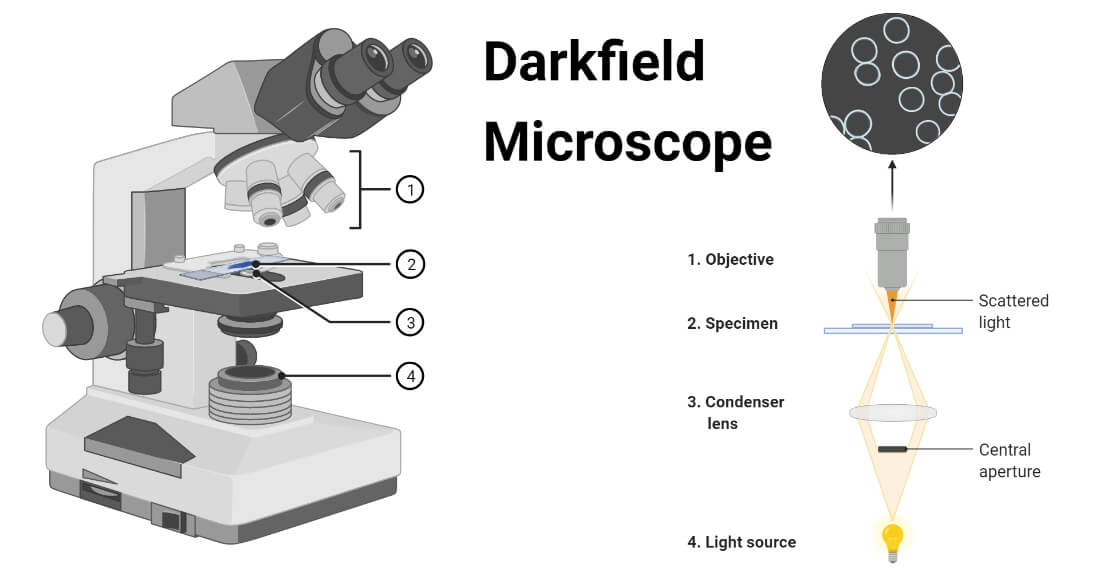
Dark Field Microscope
Principle: In a dark field microscope, the object appears bright against a dark background. This is made possible by the use of a special darkfield condenser.
Applications: It is used to identify the living, unstained cells and thin bacteria like spirochetes which cannot be visualized by light microscopy.
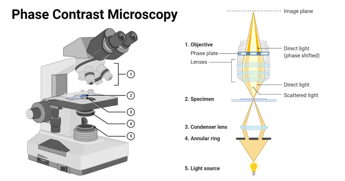
Phase Contrast Microscope
It is used to visualize the living cells by creating a difference in contrast between the cells and water. It converts slight differences in refractive index and cell density into easily detectable variations in light intensity.
It is useful for studying:
- Microbial motility
- Determining the shape of living cells
- Detecting bacterial components such as endospores and inclusion bodies.
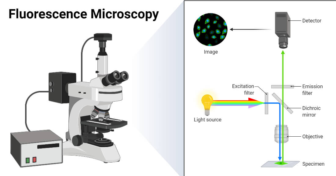
Fluorescence Microscope
Principle: When fluorescent dyes are exposed to ultraviolet rays (UV) rays, they become excited and are said to fluoresce, i.e. they convert this invisible, short-wavelength rays into the light of longer wavelengths (visible light).
Applications: Epifluorescence microscope has the following applications:
- Autofluorescence, when placed under UV lamp, e.g. Cyclospora
- Microbes coated with the fluorescent dye, e.g. Acridine orange for malaria parasites (QBC) and Auramine phenol for M. tuberculosis.
- Immunofluorescence: It uses a fluorescent dye tagged antibody to detect cell surface antigens or antibodies bound to cell surface antigens. There are three types: direct IF, indirect IF, and Flow cytometry.
Electron Microscope
It was invented by Ernst Ruska in 1931. It differs from a light microscope in various ways.
There are two types of EM:
- Transmission EM (MC type, examine the internal structure, resolution 0.5 nm, gives 2-dimensional view)
- Scanning EM (examine the surfaces, resolution 7 nm, gives 3-dimensional view)
Specimen preparation: Cells are subjected to the following steps to prepare very thin specimens (20 to 100 nm thick)
- Fixation: Cells are fixed by using glutaraldehyde or osmium tetroxide for stabilization.
- Dehydration: Specimen is then dehydrated with organic solvents (e.g. acetone or ethanol).
- Embedding: Specimen is embedded in plastic polymer and then, is hardened to form a solid block. Most plastic polymers are water-insoluble; hence complete dehydration of specimen is a must before embedding.
- Slicing: Specimen is then cut into thin slices by ultramicrotome knife, and slices are mounted on a metal slide (copper).
Freeze-etching technique: It is an alternate method for specimen preparation to visualize the internal organelles within the cells.
Cells are rapidly frozen then warmed → fractured by a knife exposing the internal organelles → subjected to sublimation → shadowed by coating with platinum and carbon.
Measures to increase the contrast of EM include:
- Staining by solutions of heavy metal salts like lead citrate and uranyl acetate
- Negative staining with heavy metals like phosphotungstic acid or uranyl acetate.
- Shadowing: Specimen is coated with a thin film of platinum or other heavy metal at a 45° angle so that the metal strikes the microorganism on only one side.


0 Comments