Cilia Definition
Cilia are tiny hair-like appendages present on the eukaryotic cell surface that provides a means of locomotion to different protozoans and animals.
- The term ‘cilia’ is a Latin term meaning eyelash indicating the tiny eyelash-like appearance of the structure.
- Cilia are most prominent in protozoans of the phylum Ciliophora which are characterized by the presence of cilia.
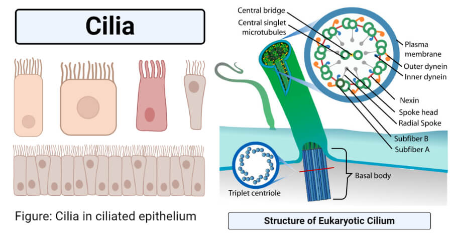
- Ciliated cells are found in different tissues in complex animals like vertebrates, where they have distinct functions.
- Cilia are different from flagella which are mostly longer and fewer in number on the cell. Cilia also differ from flagella in other aspects like composition, movement, and functions.
Read Also: 19 Differences between cilia and flagella (cilia vs flagella)

- Cilia are present only in eukaryotic cells and cannot be found on prokaryotes like bacteria. Instead, bacteria contain other structures called pili that perform similar functions to the cilia.
- On the cell surface, cilia can occur either in short transverse rows in the form of a membrane or in groups to form cirri.
- The movement of cilia mostly occurs in a rhythmic manner, and individual cilium does not move independently.
- The most essential function of cilia is movement through liquid surfaces, but in some cases, these can act as structures for mechanoreception and feeding.
- The function also differs in unicellular and multicellular organisms as in humans, the cilia present in the different epithelium are involved in moving various substances through the lumen.
- Some cells might produce immobile cytoplasmic protrusions called stereocilia which are not composed of microtubules. Stereocilia thus are considered different from true cilia called kinocilia.
- The structure and composition of cilia can be studied easily by scrapping the pharyngeal epithelium of a frog with a spatula and observing it under a microscope.
Structure of Cilium
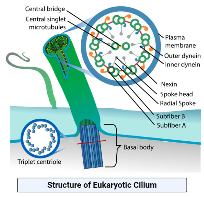
Cilia are membrane-bound, microtubule-containing, and centriole derived protrusions that project into the extracellular space. These are structurally resilient but also flexible and dynamic with distinct mechanisms to control their composition and functions. Cilia are distinguished into two types; motile cilia and nonmotile cilia, based on the patterns of microtubules present in the axonemes of the cilia. The overall basic structure of both the cilia is the same, except the axoneme.

Figure: Structure of Cilium. Image Source: LadyofHats.
The following are the parts of cilia observed in the ultrastructure;
1. Ciliary membrane
- The ciliary membrane is the outer covering of the cilia that surrounds the internal axoneme and core of the cilia.
- The membrane is continuous with the cell membrane but is different from the cell membrane in its overall composition. The membrane is about 9.5 nm thick with far fewer proteins than the cell membrane.
- Some of the proteins present in the membrane are specific to the cilia and play an essential role in preventing the loss of ATP and other ions required at specific concentrations to provide energy for the ciliary movement.
- Especially in unicellular organisms, specific receptor and channel proteins are also localized in the ciliary membrane.
- The ciliary membrane in all somatic cilia contains a region within the membrane composed of multiple strands called a cilia necklace.
- The cilia necklace acts as a selective barrier at the cilium entrance during the passage of particles from the cytoplasm.
2. Ciliary matrix
- The space within the ciliary membrane formed of a watery matrix is the ciliary matrix. The matrix consists of embedded microtubules forming the axoneme of the cilium.
3. Axoneme
- The most important structure of the cilia is the basic microtubular structure called an axoneme.
- Axoneme forms an axial structure within the cilia which is responsible for the motility of the cilia.
- The axoneme of the cilia is about 0.2-10 µm in diameter, and the length ranges from a few microns to 1-2 mm.
- Motile cilia contain an axoneme composed of a 9+2 arrangement of the microtubules. The microtubules are arranged with nine doublets surrounding a central pair of singlet microtubules.
- The microtubules are polymerized from αβ-tubulin heterodimers with a fast polymerizing end at the ciliary tip.
- The axoneme further contains outer and inner dynein arms, radial spikes, and central-pair projections. There are hundreds of proteins attached to these structures that are responsible for the assembly of the cilia as well as their function.
Cilia formation mechanism/ Ciliogenesis
- The process of formation of cilia in the cell, often termed ciliogenesis, occurs in several stages.
- Biogenesis of a cilium is a highly complex, elaborate, and regulated process occurring with the help of many organelles, cellular mechanisms, and signaling pathways.
- The formation of cilia begins after the mitotic cycle of cell division so that the free centrioles can undergo axoneme nucleation.
- The centriole during the process acquires various distal and subdistal appendages. The distal appendages interact with a post-Golgi vesicle which flattens the ciliary extension of the centriole and fuses it to the cell membrane.
- The position and orientation of the forming cilia depend on the original positioning of the basal bodies and centrioles.
- The final step of cilia formation is the formation of the actual axoneme by various molecular motors and associated proteins.
- During this step, tubulin protein subunits are assembled at the distal growing ends through the process, termed intracellular route.
- In the extracellular route, the basal body is present at the apical membrane prior to axoneme growth, and protein accumulation follows.
- The stabilization of the cilia depends on the post-translational modification of the tubulin proteins through acetylation and detyrosination.
- The proteins required for the formation of cilia are all produced in the cytoplasm and are transported to the cilia through intraflagellar transport.
- New tubulin proteins are continuously added to the cilium even after completion, but the length of the cilium doesn’t change as older tubulin begins to degrade.
- All the steps of ciliogenesis are carefully regulated by different mechanisms, and any changes in the system might affect the structure and motility of the cilia.
Types of cilia
1. Primary cilia
- Primary cilia are solitary, nonmotile cilia found in most mammalian cells that are projected from the apical surface of polarized and differentiated cells.
- Primary cilia are specialized cellular organelles like other cell organelles like mitochondria, endoplasmic reticulum and Golgi apparatus.
- Primary cilia are differentiated from other types of cilia in the presence of a 9+0 arrangement of microtubules in the axoneme.
- These lack the central singlet of microtubules which are responsible for the motility of the cilia. The cilia are anchored to the cell by means of basal body nucleated by the centriole.
- Primary cilia were discovered by Zimmerman in 1898 but were named by Sergei Sorokin in 1968.
- There are different hypotheses for the explanation of the function and structure of the primary cilia.
- The first hypothesis is that the primary cilia are vestigial organelles inherited from the ancestor with motile cilia. The second states that these are essential for the control of the cell cycle. The third hypothesis describes them as sensory organelles.
- Primary cilia are found in different cells in the mammalian body like stem cells, epithelial, endothelial, connective tissue, and muscle cells.
- Cilia that are not associated with motility are often involved in sensory functions. These cilia act as antennae that receive signals from the environment which are then converted into signaling cascades.
- These cascades begin in the ciliary compartment and are transduced to the cell body. The ciliary membrane contains various receptors, channels, and signaling proteins that are involved in the process.
- The signaling pathways employed by primary cilia are diverse and differ in different cell types.
- Aberrant form and function of primary cilia might result in disorders known as ciliopathies. These disorders have a wide range of clinical manifestations ranging from Bardet-Biedl syndrome or oral-facial-digital syndrome.
2. Motile cilia
- Motile cilia or moving cilia are cilia that are primarily involved in the movement of the organisms or different substances through a passage.
- These are typically found on the specialized epithelial lining of the airways, paranasal sinuses, oviduct, and the ventricular system of the brain.
- Motile cilia occur in large numbers and move in a coordinated beating exhibiting pendulous, unciform, infundibuliform, or undulant movement.
- Motile cilia are the only cilia that are found in ciliates that use them for locomotion or to move liquid through their surface.
- Motile cilia consist of a 9+2 structure with nine peripheral microtubule doublets and two centrally located singlet microtubules.
- The doublet microtubules consist of a complete A tubule with 13 protofilaments and an incomplete B tubule with 10 protofilaments.
- The doublets are linked together by nexin bridges responsible for the bending motions of the cilia. The doublets are connected to the central apparatus or two singlets by the means of radial spikes.
- The movement of motor cilia is regulated by optimal levels of periciliary fluid present around the cilia. The ciliary membrane of these cilia consists of sodium channels that serve as sensors to determine the fluid level around the cilia.
3. Nodal cilia
- Nodal cilia are motile cilia with a 9+0 arrangement of microtubules in the axoneme that are only present in the embryo during the early stages of development.
- Structurally, nodal cilia are similar to primary cilia except they contain dynein arms necessary for movement and spinning.
- These cilia can move in a clockwise direction resulting in the movement of extraembryonic fluid through the nodal surface.
- These cilia occur in the cells present on the node or embryonic organizer in the gastrula stage embryo.
- Nodal cilia are important for the determination of left-right orientation and movement of liquid matrix around the embryo.
- These cilia are often surrounded by primary cilia that are involved in the reception of sensory responses around the embryo.
Functions of cilia
The functions of cells might differ in different types of animals as well as in different types of cilia. The following are some of the functions of cilia;
- Cilia are the primary organ of movement or locomotion in the Phylum Ciliates of protozoans. The movement occurs as a response to changes in the environment as well as to move the extracellular fluid across the surface of the organism.
- Some organisms use the rhythmic movement of cilia to sweep away unwanted substances, other microorganisms, and mucus to prevent diseases.
- The motile cilia present in different epithelial surfaces in humans are involved in either the movement of essential substances by ovum in the oviduct or for the removal of dust particles like the cilia in the respiratory tract.
- Different cilia play important roles in cell cycle regulation and organ development.
- Nonmotile cilia or primary cilia have different proteins and receptors on the surface that act as chemoreceptors. Primary cilia in the kidney and retina are involved in the sensation of urine flow and photoreception, respectively.
- In pathogenic organisms, cilia are important virulence factors that enable colonization of different surfaces.
- In some animals, cilia have been associated with the secretion of vesicular ectosomes.
- Cilia play a crucial role in the signal transduction pathways that regulate intracellular Ca2+ level and planar cell polarity.
Examples of Cilia
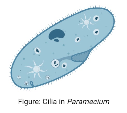
1. Cilia in Paramecium

Created with BioRender.com - Cilia are one of the most important cell organelles in Paramecium as they are involved in the locomotion of the organism through water and ingestion of food into the cytosome.
- Cilia are present throughout the body of the organism, most of which are involved in locomotion. Caudal cilia present in the gullet are usually longer and nonmotile.
- Besides, the cilia are also involved in the initial step of the mating reaction in Paramecium as it takes part in conjugation.
- The cilia sweep prey organisms along with water into the oral groove, which helps in feeding as well.
- The structure of the cilia in Paramecium is similar to that occurring in other eukaryotes consisting of a basal body, axoneme, and a ciliary membrane.
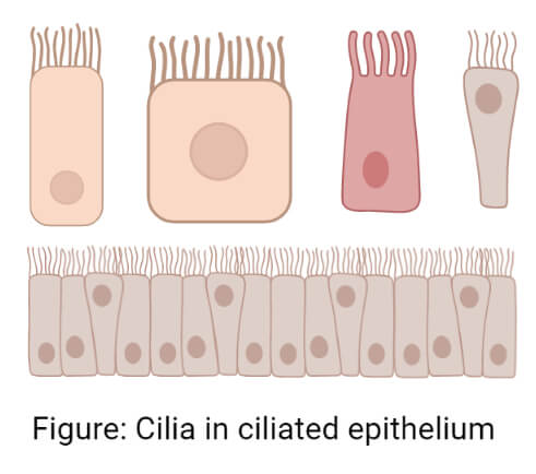
2. Cilia in ciliated epithelium

Created with BioRender.com - Cilia are present on the epithelial cells in different parts of the human body resulting in the ciliated epithelium.
- This type of epithelium commonly occurs in areas that are in close and frequent contact with the external environment.
- The ciliated epithelium of the respiratory tract is involved in the sweeping of dust particles, mucus, trapped dust, and bacteria out of the body.
- The ciliated epithelium in the retina and kidney acts as sensory structures that are involved in sensory reception.
- In the oviduct, the cilia move the ovum from the ovaries to the fallopian tubes for fertilization.


0 Comments