
Introduction
The term chromosome painting was first used by Pinkel and his group in 1988. They used this term to describe the use of complex mixtures of DNA sequences to visualise chromosome after in situ hybridization. Later a decade ago, the current technique of chromosome painting was developed independently by research teams at Lawrence National Laboratories and at Yale University. Both these groups used the cloned DNA libraries derived from flow-sorted human chromosomes.
Staining techniques like G-banding can detect large changes in chromosomes like large deletions, insertions or translocations. But they cannot detect small aberrations. Small aberrations are critical and are the cause of cancer development and progression. Sensitive mapping of chromosome aberrations across genome can be helpful in dealing small aberrations.
With the help of FISH (Florescent In Situ Hybridization) technique, individual genes or sets of repeated sequences in an intact chromosome can be detected. Later it was realized that FISH can also be used on whole genome at once. This variation of FISH technique was named as Chromosome painting. Here fluorescent dye bound DNA probe binds all along a specific chromosome.
The major drawback of FISH and chromosome painting was that, they cannot be used to study all chromosomes at the same time because there are no enough florescent dyes with sufficient color differences to mark all the 23 chromosomes of humans in a unique color. This issue was solved in 1996 by labeling painting probes for each chromosome with a different assortment of fluorescent dye called fluorochrome. The synchronized hybridization of multiple chromosome painting probes, each tagged with a specific light-emitting fluorochrome is called spectral karyotyping. When florescent probes hybridise to a chromosome, each kind of chromosome is labelled with a different variety of florescent dye combinations.
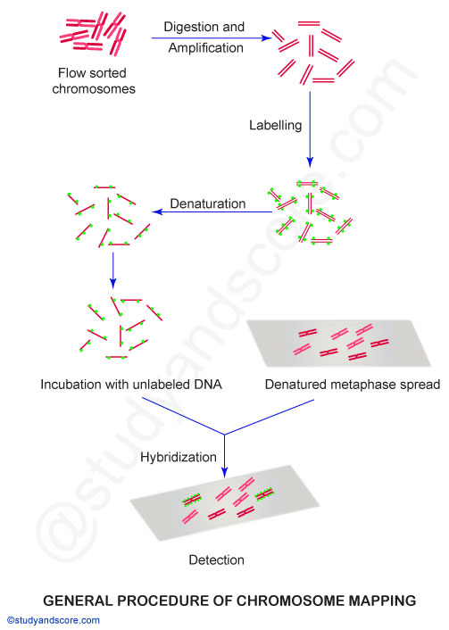
General methodology of Chromosome painting
Using these competitive hybridisation techniques, whole chromosomes can be visualised in metaphase spreads and interphase nuclei. Homologies between the chromosomes of different species can be detected by chromosome painting.
- The suspensions of chromosomes from dividing cells are sorted by flow cytometry method. The flow sorted chromosomes contain sequences which are shared with other chromosomes.
- Flow sorted chromosomes are digested and amplified with the help of Polymerase Chain Reaction (PCR)
- Then the amplified DNA from one chromosome is labelled with a fluorescent dye using a technique called fluorescence in situ hybridisation. This labelled DNA will paint and help in the identification of homologous regions of DNA of other species. We can identify the homologous regions from apes to mice. Homologous regions show up in the same colour on the chromosome charts.
- The labeled fragments are denatured again and then incubated with unlabeled DNA.
- The fluorescent-labelled DNAs will hybridise with similar chromosomes from which they were derived. Because DNA fragments with the same base sequences have the characteristic of hybridising with each other.
Technique of Chromosome painting
Chromosome painting method is actually a modification of Fluorescence In-Situ Hybridization (FISH) and is also called Zoo-FISH. Human chromosome-6 consists of hundreds of genes in major histocompatibility complex (MHC). With the help of chromosome painting researchers proved that large segments of human chromosome-6 have their homologous genes in,
- Chromosome 5 of chimpanzee
- Chromosome B2 of domestic cat
- Chromosome 7 of pig
- Chromosome 23 of cow etc.
With this technique, we can also visualise chromosome aberrations if any by using fluorescently labeled DNA probes. These labeled probes hybridize to chromosomal DNA. Even multiple fluorochromes can be attached to the probes. Upon hybridization, this produces a multicolor painted effect with a unique color at each site of hybridization. We can also detect cross-species homology by labeling probes from one species for hybridization with chromosomes from another species.
Probes used in chromosome painting
Day by day with the progress in technology, chromosome painting probes are also available for a huge number of species. Chromosome painting probes for mouse and rats are most common and are very helpful as animal models to analyse human diseases.

Chromosome painting helps in the visualization of individual chromosomes in metaphase or interphase stages. With the help of this technique, we can identify both numerical and structural chromosomal aberrations with high sensitivity and specificity. As already mentioned, the synchronized hybridization of multiple chromosome painting probes, each tagged with a specific light-emitting fluorochrome is called color or spectral karyotyping. It helps in the differential color display of human and mouse chromosomes.
Chromosome painting is also called M-FISH or multicolor FISH because modified fluorescent in situ hybridization (FISH) has been used to detect the location of specific genomic targets using probes that are labeled with specific fluorochromes. This technique allows the detection of simple and complex chromosomal rearrangements.
Applications of chromosome painting
- With the help of chromosome painting, we can get information about the divergence of lineages which has occurred over several million tears. Also, the rearrangements which took place between species can be tracked.
- Chromosome painting helps in the visualization of individual chromosomes in metaphase or interphase stages.
- Chromosome painting can identify both numerical and structural chromosomal aberrations with high sensitivity and specificity
- Chromosome painting is highly sensitive and specific.
- The application of chromosome painting in the field of comparative cytogenetics helps in understanding and determining the chromosomal changes that have occurred during the evolution of species.
- Chromosome painting identifies homologous chromosome segments in different species.
- This technique can map probes of different complexities and chromosome rearrangements in one shot-single experiment.
- In recent years, the complete karyotypes of various mammals including primates, carnivores, and artiodactyls have been analyzed by chromosome painting.


0 Comments